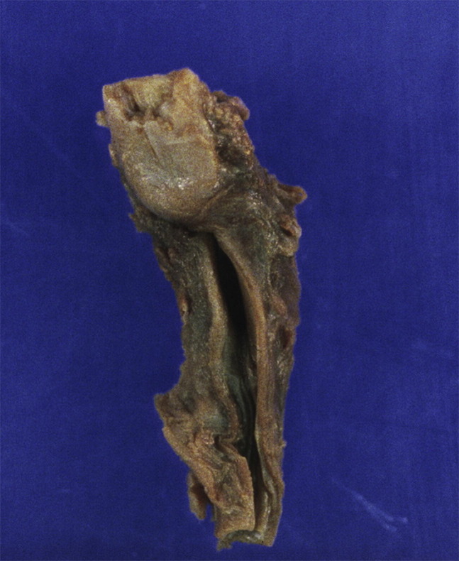
Fig. 18.
Esophageal atresia associated with extensive placental FTV. Very recent fetal death in utero occurred at 32 weeks. The largest fetal vessels at the chorionic plate appeared dilated and congested. The cord had a single umbilical artery and was tightly wrapped multiple times about the left leg of the fetus, resulting in compression marks on the leg. There were several large stem vessel thrombi; both old hyalinized villi and more recent hemorrhagic villi with extravasated red blood cells were present. Instances of atresia in the digestive tract have been attributed to vascular disruptions, most likely caused by thrombi. Unfortunately, attempts to relate bowel atresia to placental pathology are not commonly made.

