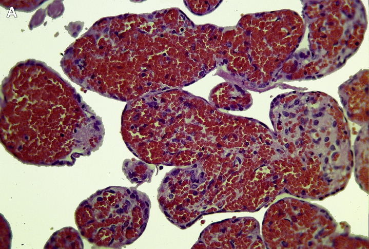
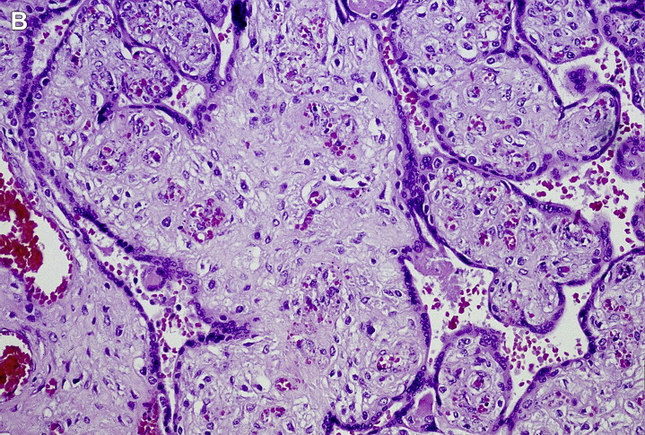
Fig. 9.
(A) Acute villous lesion. This instance of more extensive acute intravillous hemorrhage was traced to an occlusive thrombus in a large vein just before joining the umbilical vein. (B) Acute villous lesion. Fetal villous capillary lesions of recent (2–4 days) duration show disappearance of most endothelial cells, extravasation, and fragmentation of red blood cells, in a region of stasis downstream from a recent stem vessel thrombus (hematoxylin-eosin, original magnification ×300).


