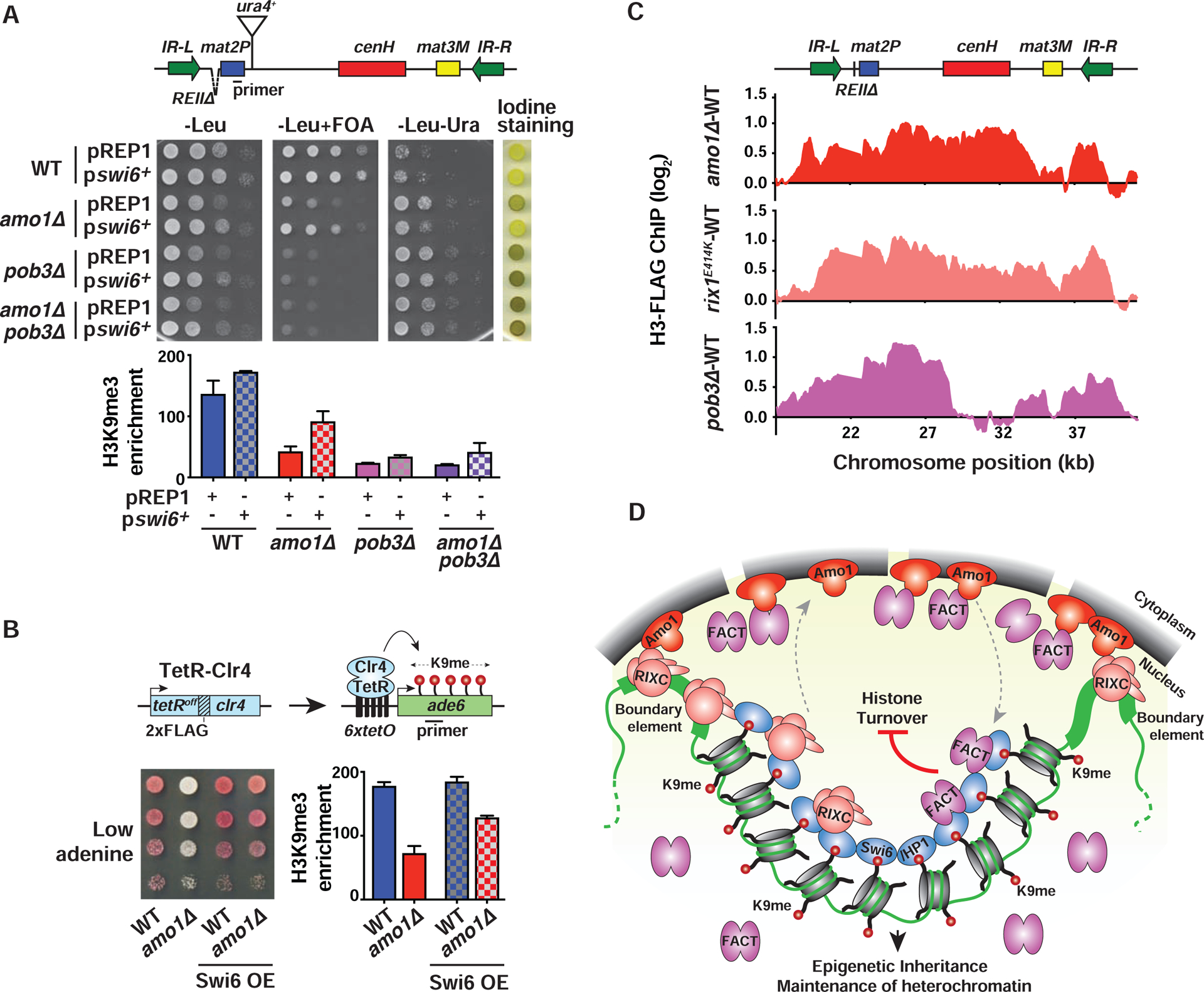
Figure 7. Nuclear peripheral positioning of heterochromatin by Amo1 suppresses histone turnover.

(A and B) Tenfold serial dilution and ChIP-qPCR analysis of H3K9me3 enrichments (mean ± SD, n=2) in WT and mutant strains transformed with LEU2-based plasmids pREP1 (control) or pswi6+ (A), or the indicated TetR-Clr4-expressing 6xtetO-ade6+ reporter strains (B). (C) Histone turnover at the mat locus as determined by H3-FLAG ChIP-chip. Strains contained REIIΔ that permits modifications affecting heterochromatin maintenance to be easily detected (see also STAR★Methods). The signals were normalized to WT. (D) Model: Amo1 positions heterochromatic loci at the nuclear periphery via RIXC and facilitates efficient loading of FACT onto Swi6HP1-bound heterochromatin. Together with HDACs (not shown), they promote epigenetic inheritance and heterochromatin maintenance by suppressing histone turnover. See also Figure S7.
