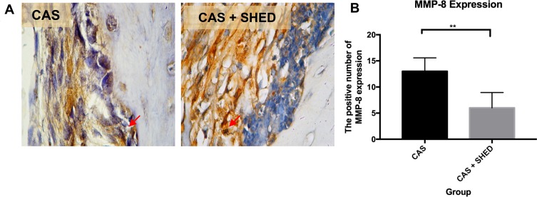
Figure 4.
The periodontal tissue histological section of an afflicted subject. (A) AbMo and DAB were used to perform immunohistochemical analysis of MMP-8 expression. Positive cells appeared brown in color (red arrow) through a light microscope at 1000x magnification. (B) The number of positive expressions of MMP-8 is shown. The statistical significance of differences between groups was examined by means of an unpaired t-test (n = 7; **Information: significant at p < 0.01).

