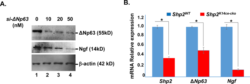
Fig.8. Down regulation of Ngf in ΔNp63 siRNA-transfected TKE2 cells and in Shp2K14ce-cko cornea.
(A) Western blotting analysis showed that Ngf protein expression level was decreased in si-∆Np63 transfected TKE2 cells. (B) qRT-PCR analysis of Shp2, ΔNp63 and Ngf mRNAs in Shp2WT and Shp2K14ce-cko corneas. Note that the 35% of Shp2 expression in the Shp2K14ce-cko was likely attributed to the contamination of non-epithelial tissue; nevertheless, both ΔNp63 and Ngf mRNAs were reduced dramatically in Shp2K14ce-cko as compared to those in Shp2WT controls. *: P< 0.05 (N=4)

