Figure 1.
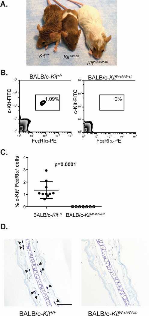
BALB/c-KitW-sh/W-sh mice are mast cell-deficient. A. BALB/c-KitW-sh/W-sh mice display a marked dilution of the agouti coat color of the wild type mice, while BALB/c-Kit+/W-sh mice display a dilution of the coat color in the characteristic “sash” pattern. B, C. Cells from peritoneal lavage were analyzed by flow cytometry for expression of the mast cell markers c-Kit and FcεRIα (B), and mast cells were quantified as a percentage of the total live peritoneal cells (C). Tissues, including ear pinnae (D), were examined for mast cells by staining with Toluidine blue. Some of the many mast cells present in the section of a wild type BALB/c-Kit+/+ mouse are indicated by solid arrowheads, whereas no mast cells are seen in this section of ear pinna from a BALB/c-KitW-sh/W-sh mice. N=7–10 mice per group for peritoneal cell analysis (C), combined from 2 independent experiments. Lines in C represent mean value ± s.e.m. Scale bars in D indicate 100 μm.

