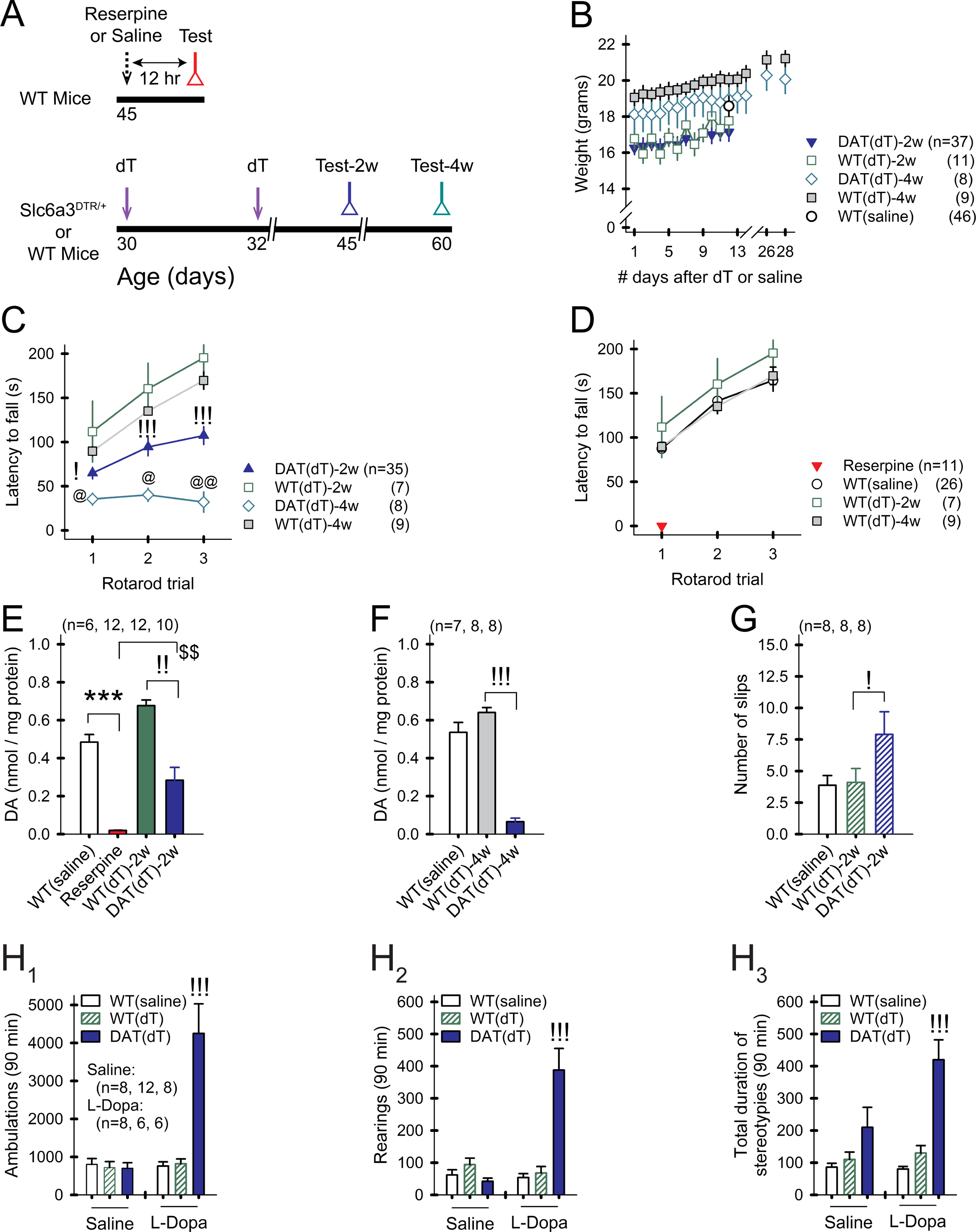
Figure 1. Progressive DA deficiency in Slc6a3DTR/+ mice.

A) Treatment timeline. B) Weights of treated mice remain stable over time. For all panels, n=mice. For all figures, data are represented as mean ± SEM; *p<0.05, **p<0.01, ***p<0.001, reserpine vs. WTsaline; !p<0.05, !!p<0.01, !!!p<0.001, DATdT vs. WTdT; @p<0.05, @@p<0.01, DATdT-2w vs. DATdT-4w; $p<0.05, $$p<0.01, $$$p<0.001, DATdT-2w vs. reserpine. C) WTsaline and WTdT mice show a progressive increase in the latency to fall from the rotarod over 3 consecutive trials. DATdT mice show a progressive reduction in rotarod performance. D) Rotarod performance is similar across control groups. Reserpine mice are incapacitated. E) Striatal DA content is depleted 12 hr after reserpine and progressively decreases 2 and F) 4 wk following the first injection of dT. G) DATdT-2w mice show impaired balance beam performance. H1) Open field tests show an increase in ambulations, H2) rearings, and H3) stereotypies in DATdT-2w mice following 5 daily treatments with L-Dopa. See also Figure S1 and Table S1.
