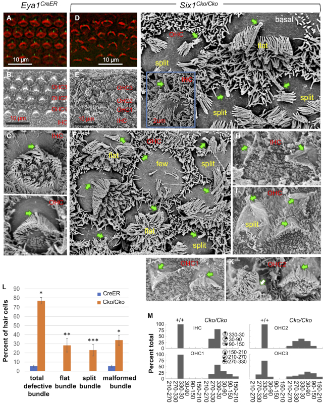
Figure 6.
Late conditional inactivation of Six1 in differentiating hair cells results in both structural polarity and PCP defects of hair-bundles. (A, D) Hair-bindle structure and orientation at P0 was visualized by F-actin (red) and anti-acetylated tubulin (green, for kinocilium). (B–K) SEM images from basal or middle cochlear duct showing surface views of the organ of Corti in Eya1CreER control and Six1Cko/Cko mutant. Arrows indicate kinocilium (C–J), which is absent in panel K. (L) Percentage of hair cells from the basal region of the cochlea of control (n = 645; 3 embryos) and Rac1 and CKO (n = 622; 3 embryos) with the indicated stereocilia. *P < 0.05, ** P < 0.01, *** P < 0.001. (M) Graphs showing distribution of hair cell orientation from wild-type and Six1 CKO mutant animals. The orientation of hair cells was determined by measuring the angle formed between the medial-to-lateral axis of the cochlea and the line bisecting the stereociliary bundle from the center of the hair cell to the vertex of the hair-bundle.

