Fig. 4.
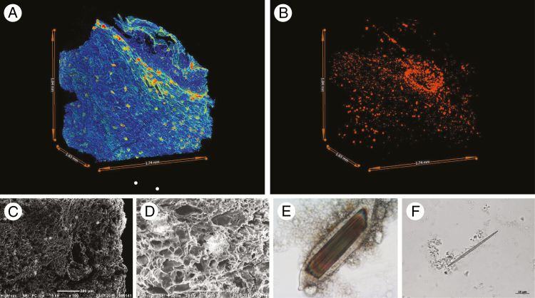
Archaeobotanical techniques for the investigation of calcium oxalate in taro (C. esculenta). (A) MicroCT visualization of a parenchyma fragment with low density areas in blue (cell walls) and high density areas in red (druses and raphide bundles comprising calcium oxalate crystals). (B) MicroCT visualization showing the distribution of high density areas in (A). (C and D) Scanning electron microscopy images of taro parenchyma with druses visible as lighter concentrations. (E) Photomicrograph of a cell packed with raphides. (F) Photomicrograph of a raphide showing needle-like morphology and an asymmetric proximal tip (lower right). All images are from modern reference samples.

