Figure 1.
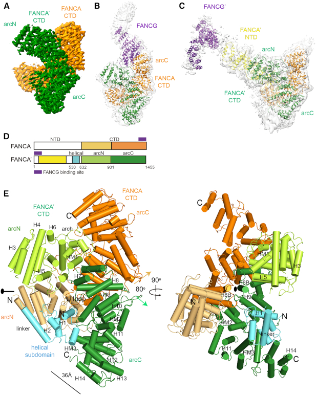
Three classes of the overall structures of the FANCA–FANCG complex. (A) Cryo-EM map of the FANCA CTD dimer at an average resolution of 3.35 Å. Each FANCA molecule is shown in orange (FANCA) and green (FANCA’). The arc-shaped CTD is divided into the N-terminal half (arcN) and the C-terminal half (arcC). (B) Cryo-EM map (gray) of the FANCA CTD dimer complexed with a FANCG (purple) molecule at 4.6 Å. FANCG CTD packs at the C-terminal end of FANCA. Orientation of the CTD dimer is same as that in (A). (C) Cryo-EM map (gray) of the FANCA and FANCG complex at 4.84 Å. FANCG’ is bound at the FANCA’ NTD (yellow). (D) Overall scheme of the subdomain composition in FANCA and FANCA’. Each subdomain is painted with same colors as in (A) to (E). (E) Two views of the 3.46 Å structure of the FANCA CTD dimer. Left, A 2-fold rotation axis is along the horizontal axis. Each HEAT repeat consists of the HA and HB helices that forms inner and outer surfaces, respectively. The central axes (along each H8B helix) of two solenoids are arranged in ∼80°. Right, View orthogonal to the left view looking down the 2-fold axis which is along the H8B helices from the two FANCA molecules. The CTD structure is divided into the N-terminal helical subdomain (H1 to linker helix), arcN (H3 to HM1) and arcC (H7 to H14) subdomains. The helical subdomain of FANCA’ is shown in cyan. The arcN and arcC subdomains of FANCA CTD are colored in light orange and orange, respectively. The arcN and arcC subdomains of FANCA’ CTD are shown in light green and green, respectively.

