Figure 3.
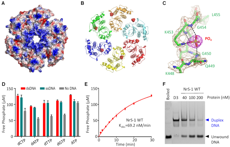
The nucleotide hydrolysis and DNA-unwinding activities of NrS-1 polymerase. (A) Surface presentation of the helicase domain of NrS-1 polymerase. Residues with positive charge and negative charge are colored in blue and red, respectively. (B) Cartoon view of the helicase domain of NrS-1 polymerase. The phosphate ions bound at the Walker A motifs are shown as spheres. (C) Detailed interactions between the phosphate group and the Walker A motif of NrS-1 helicase domain. The 2Fo− Fc electron density maps are contoured at 1.5 sigma level. (D) Analysis of the nucleotide hydrolysis activity of WT NrS-1 polymerase. (E) Time-course analysis of the dCTPase activity of NrS-1 polymerase. (F) In vitro DNA-unwinding assays catalyzed by WT NrS-1 polymerase.

