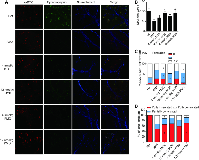
Figure 8.
NMJ staining of the flexor digitorum brevis 2 and 3 (FDB-2/3) muscles of SMA mice. (A) SMA mice were treated with saline (n = 4), MOE10-29 (4 nmol/g, n = 4; 12 nmol/g, n = 4) and PMO10-29 (4 nmol/g, n = 4; 12 nmol/g, n = 4) as in Figure 6. Untreated heterozygous mice (Het, n = 4) were used as normal controls. FDB-2/3 were collected on P9 and stained for neurofilament with anti-neurofilament (blue), nerve terminals with anti-synaptophysin (green) and motor endplates with α-bungarotoxin (α-BTX, red). (B) NMJ area was measured from (A) (four mice per group and three counts per mouse). (C) Quantification of perforations identified by α-BTX, based on (A) (4 mice per group and 3 counts per mouse). (D) Percentages of innervated endplates (red), partially denervated endplates (blue), and fully denervated endplates (white) were quantitated based on (A) (four mice per group and three counts per mouse). (*) P < 0.05 high dose versus low dose of the same ASO; (#) P < 0.05 MOE versus PMO at the same dose.

