
Figure 1.
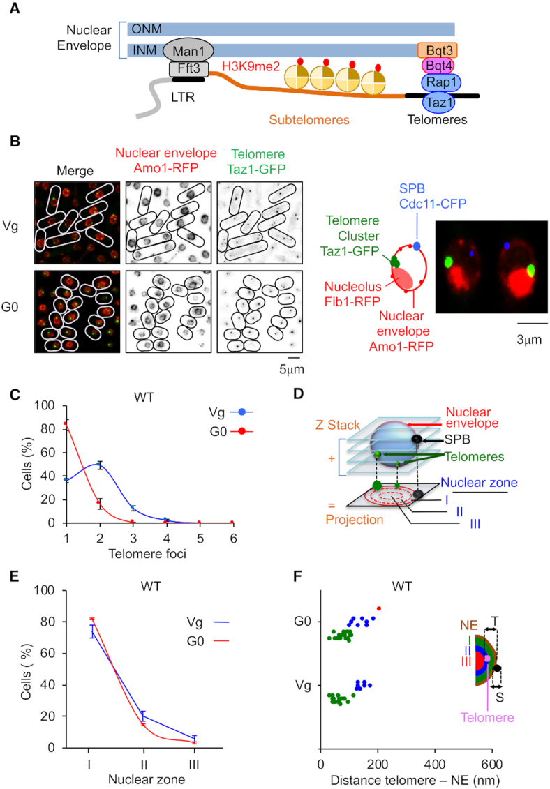
Telomeres hyperclusterize and anchor to NE in quiescent cells. (A) Schematic representation of nuclear envelope (NE) anchoring of telomeres in fission yeast. (B) Visualization of telomeric foci and NE by live microscopy in vegetative (Vg) and quiescent (G0) cells. Left panel: Taz1-GFP (green) and Amo1-RFP (red) mark the telomeres and the NE, respectively. Right panel: Image of Taz1-GFP (green), Amo1-RFP (red), Fib1-RFP (red) illustrating the Rabl conformation of telomere cluster located close to the nucleolus at the opposite of spindle pole body. (C) Percentage of cells that display one or several telomeric foci. (D) Z-stacks images of living cells and scoring of the position of the Taz1-GFP hypercluster in one of the three equal concentric zones of the nucleus with respect to Amo1-RFP in the focal plane of the GFP focus. (E) Taz1-GFP hypercluster localization relative to the NE (Amo1-RFP) in the three zones. (F) Distance to NE of the telomeric foci in Vg and G0 cells. This experiment was repeated in triplicate and for each experiment >100 nuclei with a hypercluster were analyzed. Error bars represent SEM.