Figure 6.

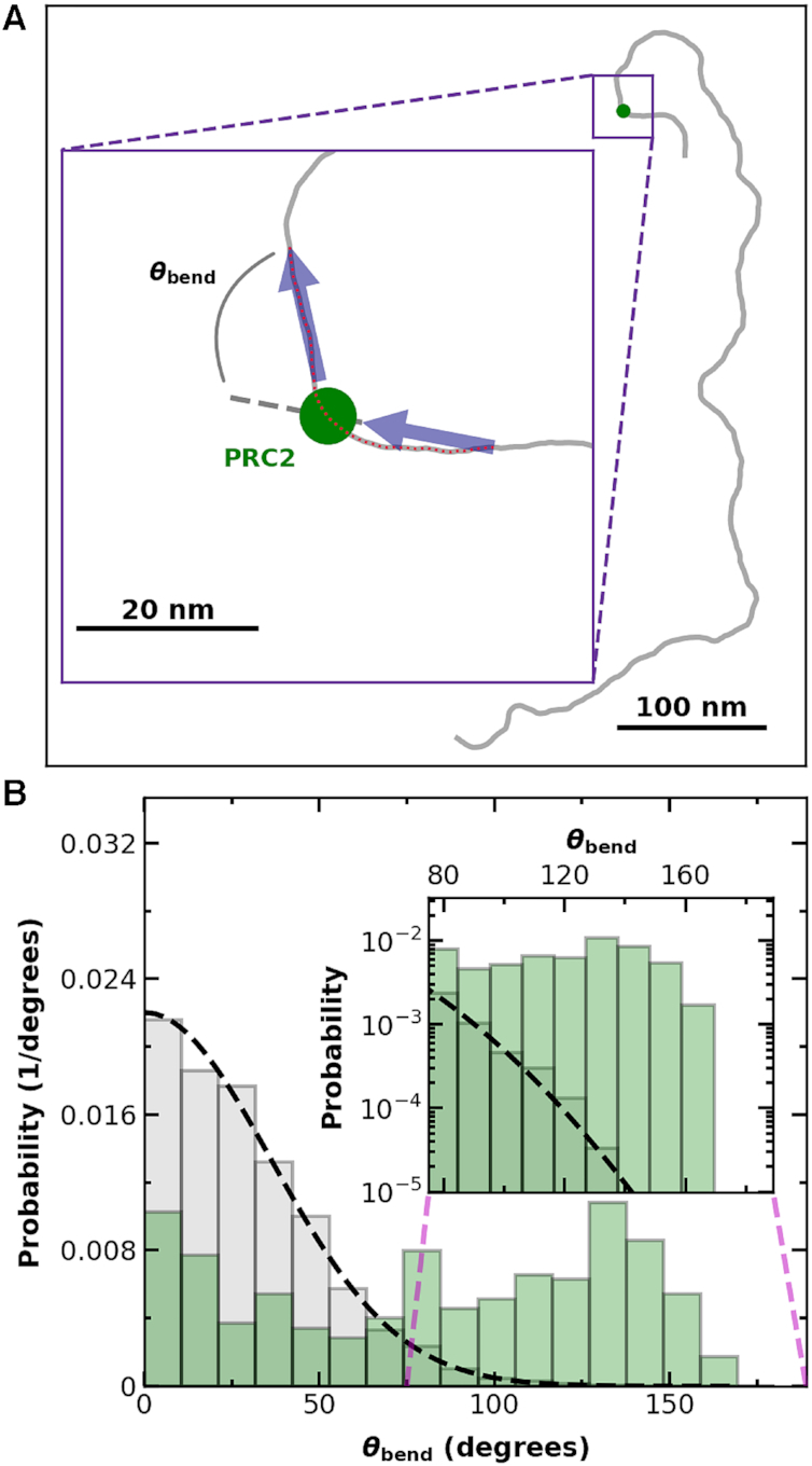
PRC2 binding is associated with an increased probability of large bends in the DNA backbone. (A) A spline denoting a 12 × 601 Widom DNA molecule (gray) bound by PRC2 (green dot). (Inset) Illustrative definition of the local bending angle (θbend) between two tangent vectors (black arrows). (B) Bending angle histogram for DNA (gray bars, Nmolecules = 70) and DNA with PRC2 bound (green bars, Nprotein = 331), where all tangent vectors were spaced 20 nm apart in contour length, and each tangent vector for the PRC2-bound DNA either started or ended at the PRC2, as shown in the cartoon in panel A. Dashed black line is the theoretically predicted bend angle distribution for tangent vectors separated by 20 nm along the DNA with a persistence length of 50 nm (54) equilibrated in two dimensions onto a surface. (Inset) Detailed distribution of large bend angle shows an excess of high angle bends for PRC2-DNA complexes in comparison to experimental and theoretical distributions of unbound DNA. Note, a single 851-nm DNA molecule gives many independent measurements of θbend.
