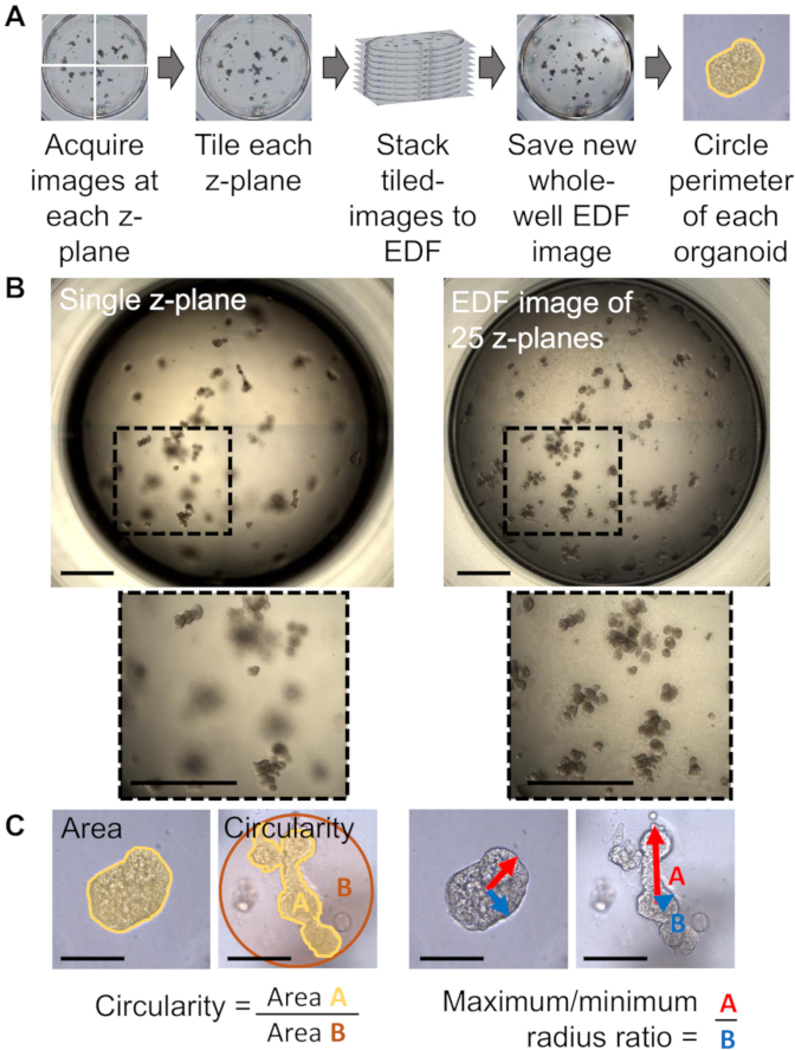
Figure 1: Morphology assessment of organoids in 3D culture.
(A) Image collection and compression workflow for human primary prostate organoids acquired by a transmitted light inverted microscope with a motorized X/Y scanning stage and companion software. (B) Representative images of a single z-stack (left) vs. an EDF image (right) of a whole-well sample of human primary prostate organoids, showing that more organoids are in focus when multiple z-stacks are combined into a single projected image (scale bar = 1000 μm). (C) Representative images for area, circularity, and max/min ratio of morphologically dissimilar human primary prostate organoids which have the same area (scale bar = 200 μm).

