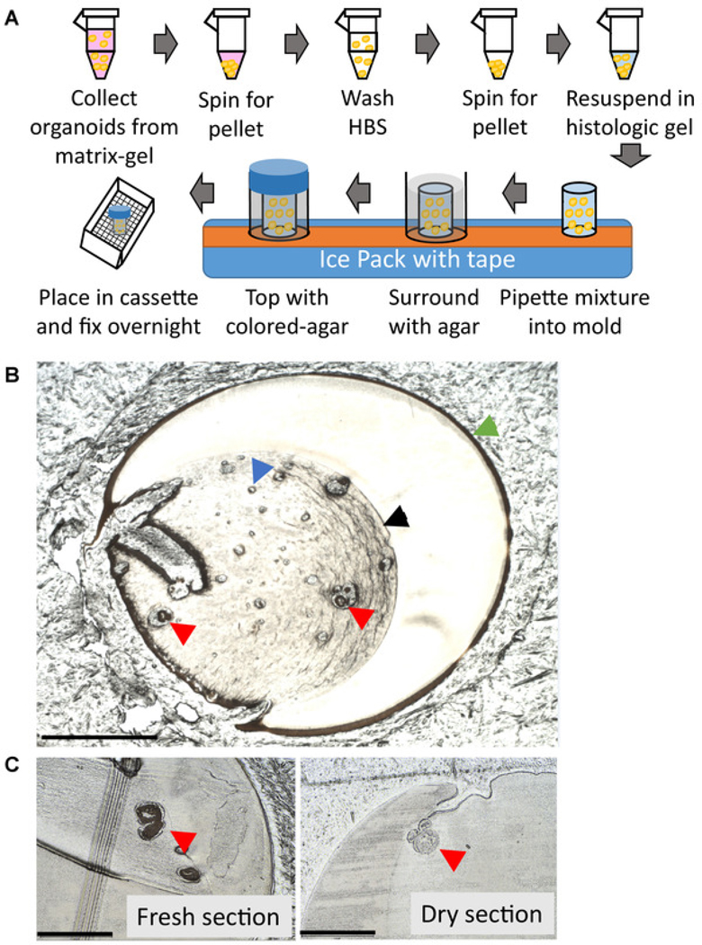
Figure 2: Organoid embedding workflow and representative sectioning results.
(A) Human primary prostate organoid embedding workflow. (B) Representative image of an unstained, fresh slide containing human primary prostate organoids under a bright field microscope depicting agar (green arrow), histology gel (black arrow), bubble (blue arrow), organoids (red arrows) (scale bar = 1000 μm). (C) Unstained freshly cut slide (left) vs. unstained dry slide (right), organoids (red arrows) appear translucent when dry (scale bar = 500 μm) and are harder to discern under a bright-field microscope.

