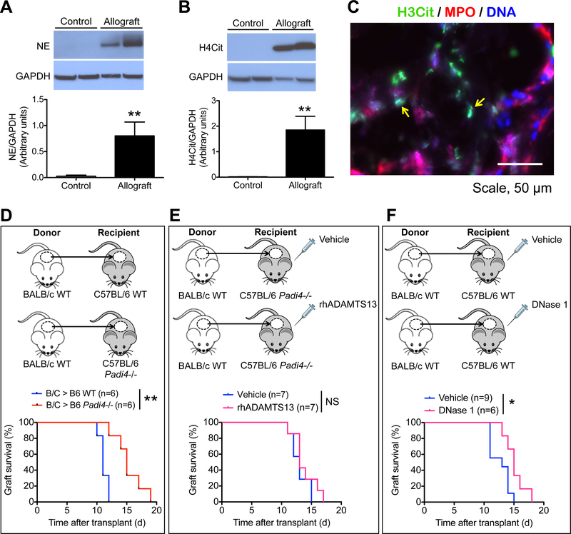
Figure 2.
NETs are present in the allografts of burn patients, and anti-NET strategies promote allograft survival in mice. (A, B) Representative Western blots and summarized data of the levels of (A) neutrophil elastase (NE) and (B) H4Cit in control skin (intact skin adjacent to burn wounds or skin collected for autografts) and allografts from burn patients. n = 4 for control, n = 6 for allograft, **P<0.01, Mann-Whitney test. (C) Representative immunofluorescence image showing the presence of NETs (yellow arrows) in the allograft of a burn patient. (D-F) Allograft survival curves of (D) WT versus Padi4−/− mouse recipients, (E) Padi4−/− allograft recipients treated with vehicle or rhADAMTS13, and (F) WT allograft recipients treated with vehicle or DNase 1. n = 6–9 per group as indicated on the graphs; *P<0.05, **P<0.01, NS, non-significant, Mann-Whitney test.

