Figure 2:
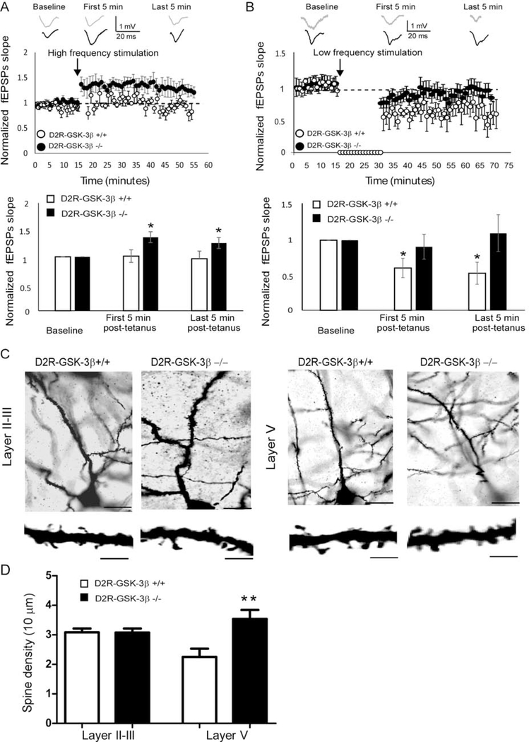
Neuronal plasticity and dendritic spine were altered in D2R-GSK-3β−/− mice. A, LTP induction. Upper: representative fEPSP traces were recorded from mPFC layer V neurons of D2R-GSK-3β+/+ mice and D2R-GSK-3β−/− mice and graphical representation showing normalized fEPSP slope during baseline recording and following high-frequency (six trains of 100 pulse at 100 Hz) stimulation. Lower: summary histogram showed that the fEPSP slopes in both first 5 min and last 5 min after high-frequency stimulation were significantly increased in D2R-GSK-3β−/− mice, but not in D2R-GSK-3β+/+ mice, suggesting an increased LTP induction in D2R-GSK-3β−/− mice (n = 10, D2R-GSK-3β+/+: p > 0.05 for both the first and last 5 min post-tetanus; n = 10, D2R-GSK-3β−/−: * p < 0.05, both the first and last 5 min post-tetanus). B, LTD induction. Upper: representative fEPSP traces were recorded from mPFC layer V pyramidal neurons of D2R-GSK-3β+/+ mice and D2R-GSK-3β−/− mice and graphical representation showing normalized fEPSP slope during baseline recording and following low-frequency stimulation (900 pulses at 1Hz). Lower: summary histogram showed that the fEPSP slopes in both the first 5 min and last 5 min of low-frequency stimulation were decreased in wild-type D2R-GSK-3β+/+ mice, but the no LTD was induced in D2R-GSK-3β−/− mice, suggesting a terminated LTD in D2R-GSK-3β−/− mice (n = 8, D2R-GSK-3β+/+: *p < 0.05 for both the first and last 5 min post-tetanus; n = 8, D2R-GSK-3β−/−: p > 0.05, both the first and last 5 min post-tetanus). C, upper: Golgi–Cox-stained individual layers II-III and layer V pyramidal neurons in the mPFC from D2R-GSK-3β+/+ and D2R-GSK-3β−/− mice. High-magnification images of apical dendritic spines were shown in the lower panels. Scale bars = 50 μm for the upper panel and 10 μm for the lower panel. D, summary histogram showed that spine density in layer II-III and layer V pyramidal neurons of D2R-GSK-3β+/+ and D2R-GSK-3β−/− mice (n = 10 from 4 mice for each group, p > 0.05 for layer II-III and *p < 0.05 for layer V).

