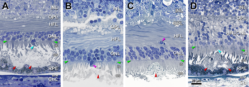
Figure 3. Solitary SDD protrude into overlying inner segments.
INL, inner nuclear layer; OPL, outer plexiform layer; HFL, Henle fiber layer; ONL, outer nuclear layer; IS, inner segment; OS, outer segment; RPE, retinal pigment epithelium; BrM, Bruch membrane; Ch, choroid; Green arrowheads, ELM, external limiting membrane. Red arrowheads, SDD, subretinal drusenoid deposits. Scale bar in (D) applies to all panels. SDD are artifactually split in B,C. A. 1400 μm temporal. A large SDD indents the apical surface of RPE. Photoreceptor OS fragments form a cap on the SDD surface, and overlying IS are short (teal arrowhead). Eighty-five-year-old woman. B. 1800 μm nasal. SDD fragments, absent photoreceptor OS, and short IS, one with an ectopic nucleus (purple arrowhead). Eighty-five-year-old woman. C. 850 μm nasal. SDD bulges into photoreceptor IS. Deposit contains heterogeneous debris including a few melanosomes. Photoreceptors OS disappear and IS are short over the deposit apex. Ectopic photoreceptor nuclei (an early indication of dyslamination) localize to the HFL (purple arrowhead). Eighty-eight-year-old man. D. 1900 μm nasal. SDD mound makes wavy concavities in the RPE surface that result in a thinned but still intact layer. Photoreceptor OS, recognized by presence of disks, are found within SDD, underlying short IS (teal arrowhead). Dyslamination of HFL and ONL is apparent. Seventy-six-year-old woman.

