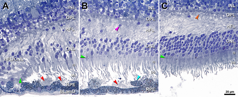
Figure 4. Border of geographic atrophy with subretinal drusenoid deposits.
INL, inner nuclear layer; OPL, outer plexiform layer; HFL, Henle fiber layer; ONL, outer nuclear layer; IS, inner segment; OS, outer segment; RPE, retinal pigment epithelium; BLamD, basal laminar deposit; Green arrowheads, ELM, external limiting membrane. Red arrowheads, SDD, subretinal drusenoid deposits; Scale bar in (C) applies to all panels. A. 1500 μm temporal. The ELM descent is a curved line that delineates the border of atrophy signified by OS absence and IS shortening at the descent. SDD are set back from this border on a wavy but intact RPE layer. The HFL and ONL is dyslaminate, and the HFL is disordered. B. 150 μm temporal to the ELM descent, SDD are observed, with sloughed RPE (teal arrowhead) and longer photoreceptors than in A. In the overlying ONL and HFL are several ectopic nuclei (purple arrowhead). C. 400 μm temporal to ELM descent, the HFL is disordered with only one ectopic nucleus. Cone pedicles with dark staining synaptic complexes (orange arrowhead) indicate good tissue fixation quality. OS were artifactually detached from the RPE. Seventy-six-year-old woman.

