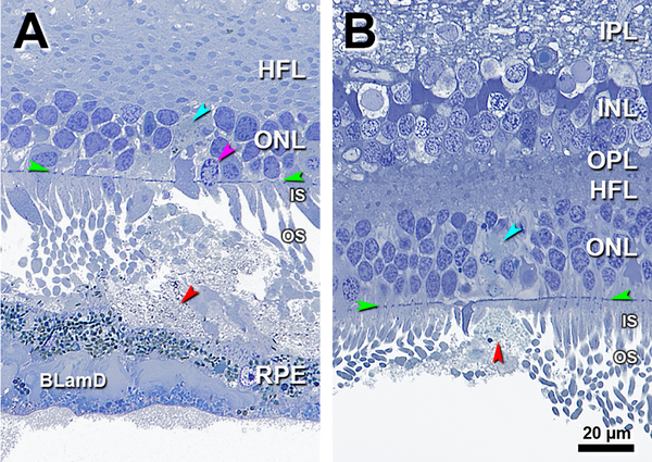
Figure 6. Subretinal drusenoid deposit material in the ONL.
IPL, inner plexiform layer; INL, inner nuclear layer; OPL, outer plexiform layer; HFL, Henle fiber layer; ONL, outer nuclear layer; IS, inner segment; OS, outer segment; RPE, retinal pigment epithelium; BrM, Bruch membrane; Ch, choroid; BLamD, basal laminar deposits. Green arrowheads, ELM, external limiting membrane. Red arrowheads, SDD, subretinal drusenoid deposits; Scale bar in (B) applies to all panels. A. 1300 μm temporal. SDD materials are interspersed among photoreceptors with an ONL gap (teal arrowhead) possibly indicating SDD material or disorganized cell processes. Photoreceptors IS are shortened and deflected, and one photoreceptor nucleus is displaced to IS (purple arrowhead). RPE with thick BLamD overlies soft druse with partial contents due to detachment. Eighty-five-year-old woman. B. 2500 μm temporal. SDD closely approximates the ELM, and a blue stained gap in the ONL (teal arrowhead) may represent SDD material or disorganized cell processes. OS disappear and IS are very short. The SDD apex detached from the base. Eighty-three-year-old woman.

