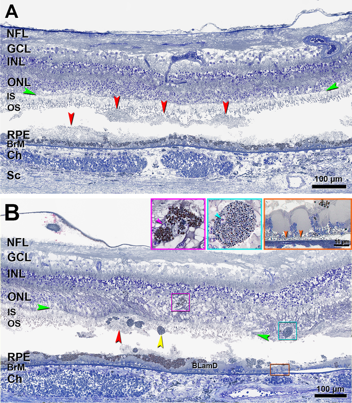
Figure 9. Histology of stages 2–3 subretinal drusenoid deposit in an eye with geographic atrophy and incipient outer retinal atrophy.
Histology comes from the patient in Figure 8. NFL, nerve fiber layer; GCL, ganglion cell layer; INL, inner nuclear layer; ONL, outer nuclear layer; IS, inner segment; OS, outer segment; RPE, retinal pigment epithelium; BrM, Bruch membrane; Ch, choroid; Sc, sclera; BLamD, basal laminar deposit; Green arrowheads, ELM, external limiting membrane. Red arrowheads, SDD, subretinal drusenoid deposits; Scale bar in the aqua inset also applies to the purple inset. In both panels, SDD split and contents were partially lost. A. The undulating nature of these SDD is well visualized. Photoreceptor OS are affected but IS are intact (stage 2 SDD). B. In the same region as the orange frame in Figure 8F, SDD fragments can be observed in the sub-retinal space. Photoreceptor OS disappear, IS are very short, sloughed RPE (yellow arrowhead) is present exactly at the stage 3 SDD. Two intraretinal RPE cells with different shapes and internal contents can be observed (purple and aqua frames and insets). The purple inset shows the same cell in another section in this series. This cell has an irregular shape and a small nuclear profile (arrowhead); pigment granules inside this cell are similar in size and color to cells in the RPE layer. The cell in the aqua inset is ovoid, and it has a small nucleus (arrowhead) and its pigment granules are smaller and less densely packed than those in the RPE layer. At the right is geographic atrophy (Figure 8C,F), with thick BLamD, and delimited by an ELM descent (green arrowheads). Of the atrophic area, a 300 μm length is shown, and 110 μm is off the right edge. BLinD is a gray thin layer continuous with an area of RPE-BLamD detachment from Bruch’s membrane (orange frame and inset, orange arrowheads).

