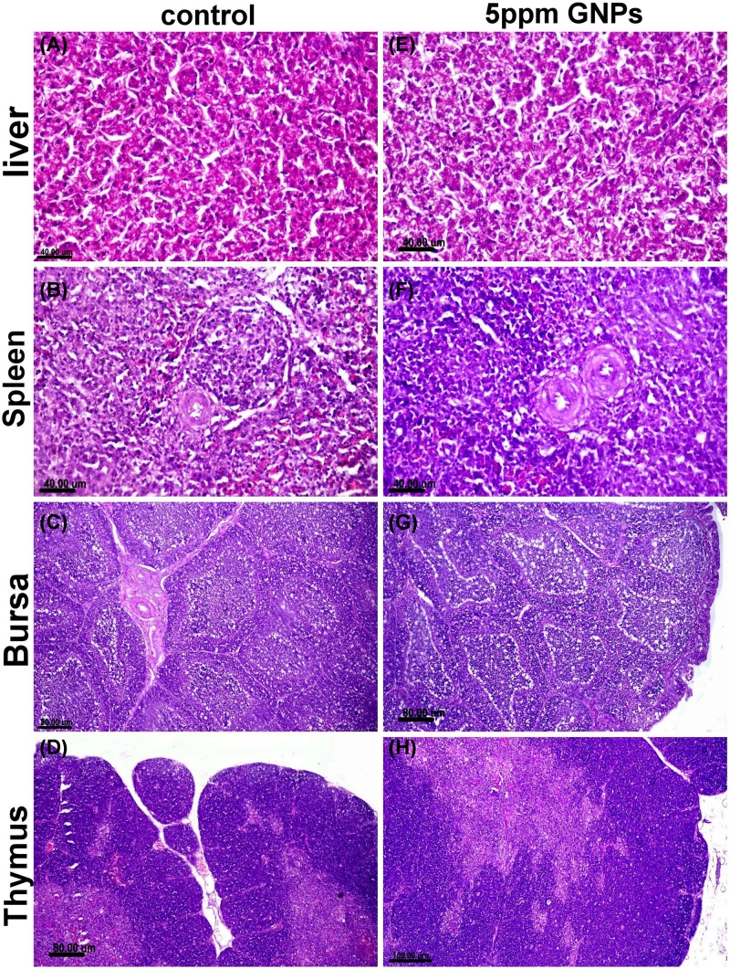
Figure 4. Histopathological examination of different organs from groups recieved 5 ppm GNPs.
Photomicrograph of liver, spleen, bursa of fabricius and thymus tissue sections stained with (H,E) of control negative group (A–D) and those receiving 5 ppm GNPs (E–H) showed normal histology with minimum pathological alteration in group receiving 5 ppm GNPs.

