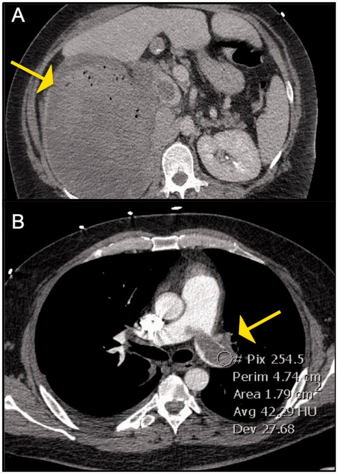
Fig. 11.
Panel A: Axial image from CT abdomen showing a large heterogenous right renal mass which was found to be renal cell carcinoma Panel B: Corresponding image from CT-PE protocol exam showing an enhancing filling defect in the left main PA. This was surgically removed and found to be a tumor thrombus.

