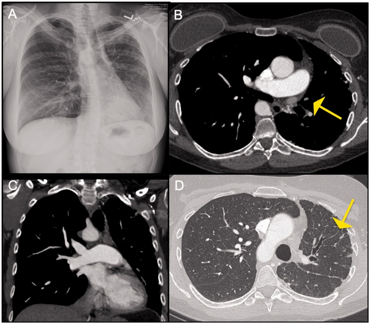
Fig. 12.
Panel A: CXR showing mediastinal shift to the left with volume loss in a patient with congenital interruption of the left pulmonary artery. Also noted is shallow appearance of the left hilum. Panel B and C: Axial CT images in mediastinal window settings demonstrate complete absence of the left PA. Few bronchial artery collaterals are noted in the left hilar region. Panel D: Corresponding lung window image shows volume loss of the left lung with few peripheral reticulations (which reflect nonspecific fibrosis).

