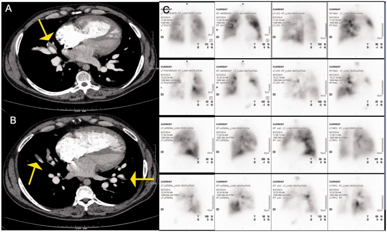
Fig. 2.
Panel A and B: Contrast enhanced CT images in a 50-year-old patient with in situ thrombosis in Eisenmenger's syndrome with an unrepaired atrial septal defect demonstrating multiple eccentric filling defects. Unlike typical webs of CTEPH, these are fairly smooth in appearance and the vessel appears normal in caliber. Panel C: Planar ventilation and perfusion images in multiple projections in the same patient. The perfusion images are more homogenous than the ventilation images in general. Multiple matched defects are noted in both lungs such as in the anterior segment of the RUL, RML, Superior segment of the RLL, Anterior segment of LUL, superior segment of the LLLL, and the posteromedial segment of LLL, No segmental mismatched defects are seen in either lungs to suggest CTEPH.

