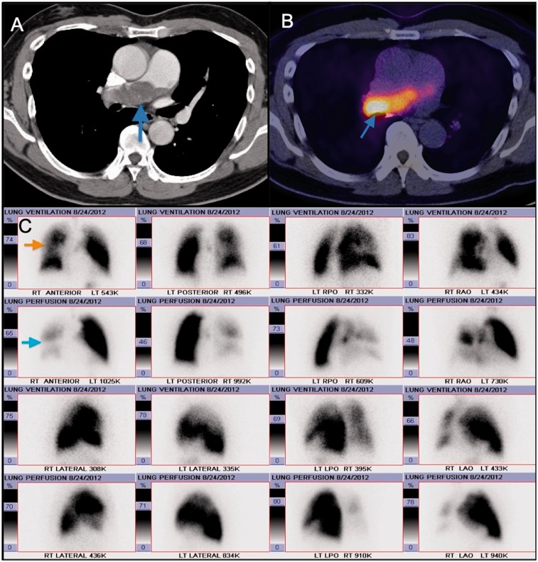
Fig. 3.
Panel A and B: Axial contrast enhanced CT mage showing a large filling defect with a lobulated appearance in the main pulmonary artery extending to the right main PA. Corresponding fused PET-CT image showing FDG uptake in the lobulated mass in the right main pulmonary artery, consistent with neoplastic process. Panel C: Images from corresponding planar ventilation-perfusion scan demonstrate mismatched perfusion defects in the right lung with severely compromised perfusion.

