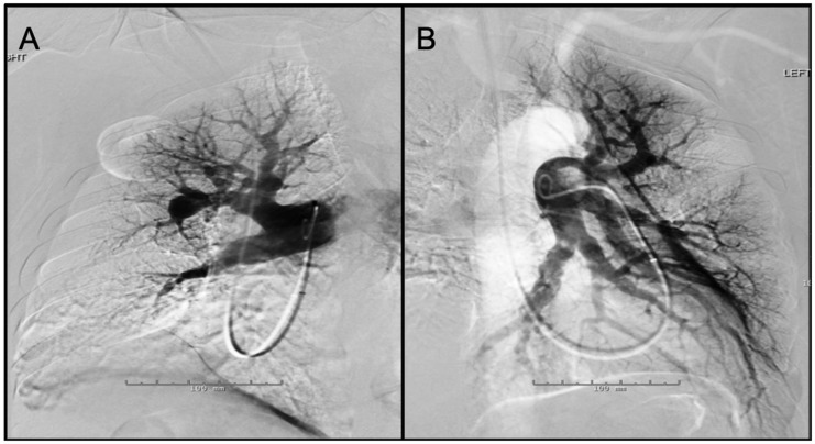
Fig. 7.
Panel A: Digital subtraction image from a right pulmonary angiogram in LAO 42 ° projection – There is stenosis of anterior segmental branch of RUL with post-stenotic dilation. Also noted is atrophic basilar segmental branch of the RLL. The perfusion in the periphery of the RUL (anterior segment) is diminished. Panel B: Digital subtraction image from a left pulmonary angiogram in RAO 37 ° Caudal 1 projection – There are atrophic/occluded lingular segmental branches. The basilar segmental branch of the LLL demonstrates luminal irregularities without significant luminal narrowing. There is associated decreased perfusion of the lingula.

