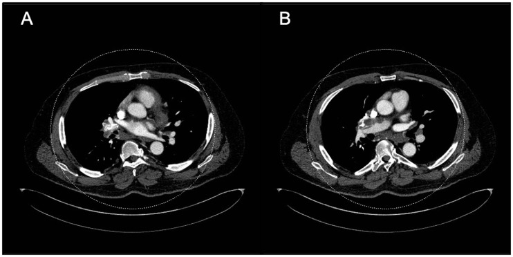
Fig. 9.
Axial CT images in the mediastinal window settings demonstrate calcified confluent mediastinal and right hilar lymphadenopathy in a patient with combined CTEPH and sarcoidosis. Also noted is lobular filling defects in the right main and interlobular pulmonary artery, consistent with CTED; there is mass effect on the right interlobar PA secondary to the calcified lymphadenopathy.

