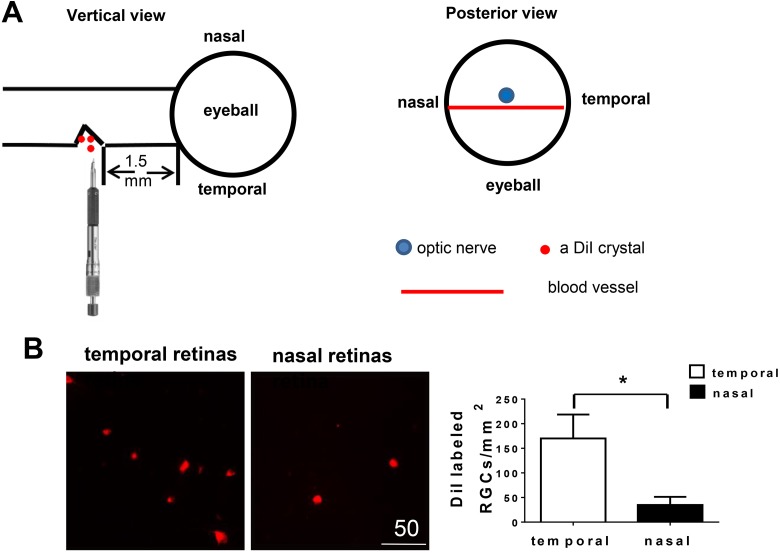
Fig. 1.
Partial optic nerve transection (PONT) model from the temporal side of optic nerves. (A) Schematic diagram showing the cut site of optic nerve and the structure (blood vessel) assisting the localization of the cut site during the surgery of PONT. After PONT, several crystals of DiI were immediately put into the cut site. (B) Retinal ganglion cells (RGCs) labeled with DiI in vivo. Photographs about 1.5 mm from the optic disc showed the different densities of DiI labeled RGCs in both the temporal and nasal retinas. More RGCs in the temporal retinas were labeled with DiI than in the nasal retinas 4 days after PONT (n = 6, Mean ± SEM, Student pair t-test, *p < 0.05).

