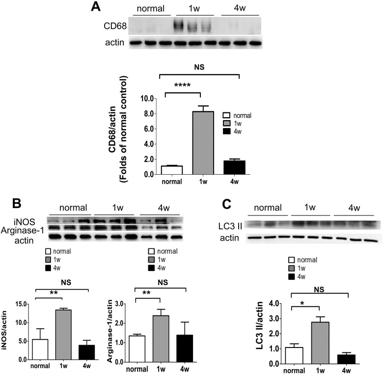
Fig. 3.
Examination of the changes in microglia/macrophages and autophagy in optic nerves after PONT. (A) Effects of PONT on the activation of microglia/macrophages. Western blot analysis shows that the expression level of CD68 increases significantly 1 week after PONT in the injured optic nerves (student t-test, ***p < 0.001), but there was no significant difference between optic nerves from rats 4 weeks after PONT and that of normal retinas (student t-test, p > 0.05). (B) Effects of PONT on the polarization of microglia/macrophages. The expression levels of iNOS and Arginase-1 increase significantly 1 week after PONT (Student t-test, **p < 0.01); but not 4 weeks after PONT (Student t-test, p > 0.05). (C) Activation of autophagy after PONT. The LC3 II expression increased significantly 1 week after PONT (Student t-test, *p < 0.05); but not 4 weeks after PONT (Student t-test, p > 0.05). (mean ± SEM; NS: not significant; n = 6 in each group for Western blot analysis, two optic nerves in one sample.).

