Fig. 4.
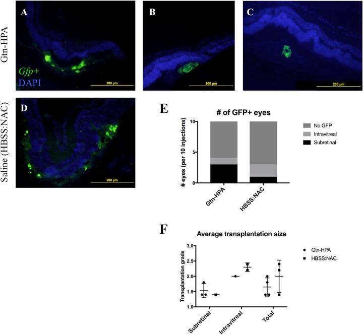
Transplantation of GFP+ pRPCs in immunosuppressed Long Evans rats. Specimens were obtained 168 hours after transplant. (A–C) Examples of subretinally transplanted GFP+ cells with Gtn-HPA. Top of image = Vitreous side, Bottom = Choroidal side. GFP+ cells are below the outer nuclear layer (DAPI positive). (D) Example of subretinally transplanted GFP+ cells in saline control (HBSS-NAC). Looser distribution of GFP signal compared with A–C. No migration or integration into inner layers of retina seen with Gtn-HPA nor saline control. (E) Number of injected eyes with GFP+ findings, per 10 injections. Gtn-HPA showed more subretinally remaining transplants. A number of eyes showed GFP+ cells intravitreally in both groups, despite a confirmed subretinal bleb formation during transplantation. (F) Average transplantation grade of GFP+ eyes, calculated as average of five consecutive sections with the best grade (grade 3: >100 cells, grade 2: 10-100 cells, grade 1: 1–10 cells, grade 0: 0 cells per section). No significant difference seen between Gtn-HPA and HBSS-NAC groups.

