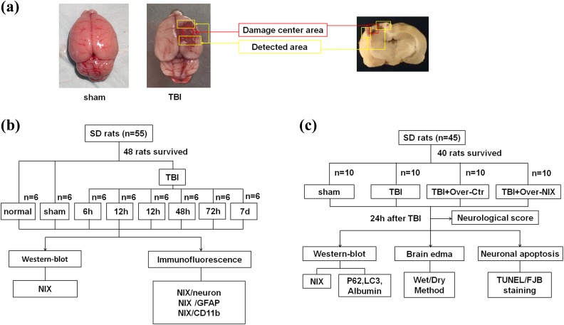
Fig 1.
The experimental process. a. Detected areas of brain tissues in the TBI group and the same location in the sham group were showed as the figure, which also indicated in the coronal sections of the damaged brain. b. Experiment I was designed to observe the expression of NIX at different time points after TBI and to find a specific time point for the next experiment. Furthermore, the location of NIX in brain cells was also confirmed at this time point. c. Experiment II was designed to investigate the possible effects of NIX on TBI-induced damage.

