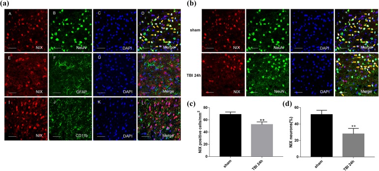
Fig 3.
The location of NIX in the cortex of the sham group and changes at 24 h after TBI by double immunofluorescence staining. a. Sections from the sham group were labeled with NIX (red A, E, I), different cell markers (green B, F, J) such as a neuronal marker (NeuN), astrocyte marker (GFAP), and microglial marker (CD11b), as well as DAPI (blue C, G, K) to mark the nucleus. In the merged images (D, H, L), cells shown by the orange arrows could be found in neurons on the whole, but not in astrocytes or microglial cells. b. NIX-positive neurons in the peri-injury cortex at 24 h after TBI were also observed and compared with those in the sham group. c. Quantification of the number of NIX positive cells/mm2 in TBI group and sham group. The results showed that NIX-positive cells were significantly decreased at 24 h after TBI. d. Quantitative analysis was used to verify the differences of NIX-positive neurons between these two groups. Scale bars, 50 µm. Data are shown as the mean±SEM (n = 6, **P < 0.01 vs sham).

