Figure 3.
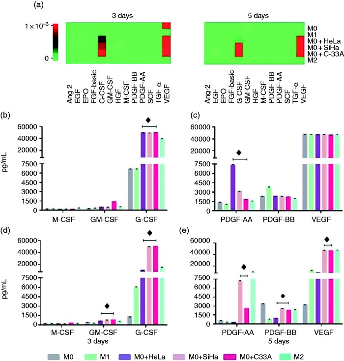
Secretion of VEGF, PDGF, GM-CSF, and G-CSF by macrophages is increased by the addition of HeLa, SiHa, and C-33A supernatants. Macrophages were obtained by differentiation of the U937 cell line with PMA (M0 macrophages), stimulation with LPS (M1 macrophages) treatment with IL-10 (M2 macrophages), or incubation in the presence of 30% of HeLa, SiHa, or C-33A for 3 and 5 d (see Materials and methods). Growth factors were quantified using bead-based multiplex assay by flow cytometry. (a) Heatmaps generated by LEGENDplex™ Data Analysis software, representing changes in the concentrations of growth factors at d 3 and 5 for each experimental group. (b)–(e) The most representative growth factors were graphed based on the corresponding analyses for different macrophages. Results of each experimental condition were obtained from assays conducted in triplicate and are represented as the mean ± SD of concentrations in pg/ml. ♦p < 0.05 HeLa, SiHa, and C-33A vs M0 and M1; ● p < 0.05 HeLa, SiHa, and C-33 vs M1; ♦ p < 0.05 SiHa and C-33A vs M0 and M1.

