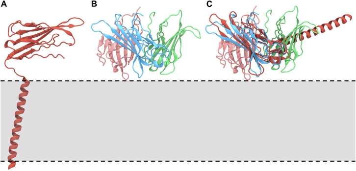
FIGURE 1.
Orientations of the β1/3 subunit on the membrane. (A) The Nav β1 subunit complex, highlighting the orientation, and interaction of the β1 subunit with respect to the membrane [PDB: 6AGF (Pan et al., 2018)]. (B) Structure of the trimeric Ig domain from β3 [PDB: 4L1D (Namadurai et al., 2014)]. (C) Overlay of the trimeric β3 Ig domain on the Ig domain of the β1 subunit, demonstrating the anticipated position of the β1 TMD and suggesting that these conformations are not compatible. The approximate location of the membrane is indicated by a gray box and dotted lines.

