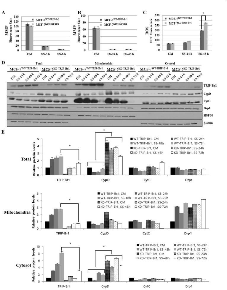
Fig. 3. Effect of TRIP-Br1 on mitochondrial functions (MMP and ROS generation) and the release of necroptosis inducer (CypD) in response to serum starvation.
Stable MCF-7WT-TRIP-Br1 and MCF-7KD-TRIP-Br1 cell lines were incubated in media with or without serum for the indicated durations. (A and B) MMP was tested as described in Materials and Methods section after MCF-7WT-TRIP-Br1 and MCF-7KD-TRIP-Br1 cells were cultured in complete media (CM) or serum starved (SS) media under two different conditions: short (3 h and 6 h), and long (24 h and 48 h) exposure to serum starvation. Data are presented as mean ± SD based on three independent experiments. *P < 0.05. (C) Cellular ROS levels were measured as described in Materials and Methods section after culturing MCF-7WT-TRIP-Br1 and MCF-7KD-TRIP-Br1 cells in complete media (CM) and serum starved (SS) media for 24 h and 48 h. Intracellular ROS levels were evaluated using 2’, 7’-dichlorodihydrofluorescein. Data are presented as mean ± SD based on three independent experiments. *P < 0.05. DCF, dichlorofluorescein. (D) Stable MCF-7WT-TRIP-Br1 and MCF-7KD-TRIP-Br1 cell lines were cultured in either complete media (CM) or serum starved medium (SS) for 24, 48, and 72 h followed by mitochondrial fractionation as described in Materials and Methods section. Isolated protein fractions were analyzed using western blot for protein expression. HSP60 was used as a mitochondrial marker and β-actin as a cytosolic fractionation marker. (E) Results of western blot were quantified using ImageJ program. Data are presented as mean ± SD based on three independent experiments. *P < 0.05.

