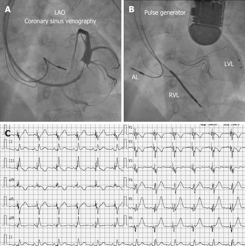
Figure 5.
Coronary sinus venography and electrocardiogram. A: Coronary sinus venography performed from a left anterior oblique 30° angle, and presentation of the coronary vein; B: Anteroposterior view of the position of the final leads; C: Twelve-lead electrocardiograms revealed biventricular pacing and a QRS duration of 104 ms after successful implantation of a cardiac resynchronization therapy-defibrillator. AL: Atrial lead; RVL: Right ventricular lead; LVL: Left ventricular lead. LAO: Left anterior oblique.

