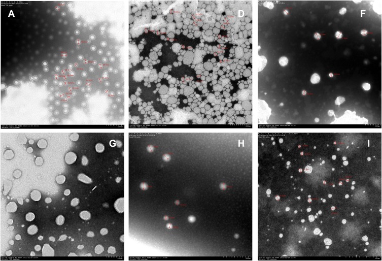
Figure 2.
Electron microscopy of nanoemulsions (Each nanoemulsion sample was diluted with an appropriate amount of PBS, added with glycerin and ultrasonically dispersed, added to a copper mesh to be dried, and stained with phosphotungstate). Magnification: (A×12,000; D×15,000; F×12,000; G×12,000; H×8000; I×25,000).

