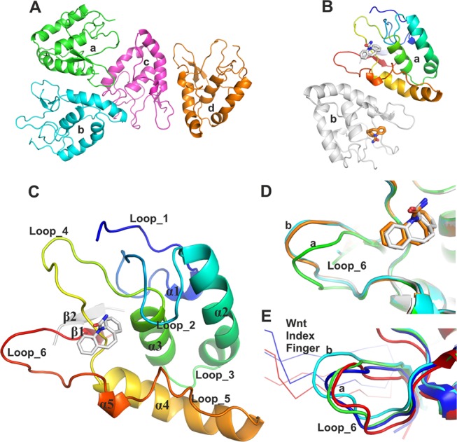
Figure 2.

Overall structure of apo and carbamazepine-bound FZD8CRD and structural comparison. (A) Four molecules of the apo structure of FZD8CRD in the asymmetric unit (ASU) (PDB code 6TFM). (B) Two molecules of FZD8CRD in complex with carbamazepine in the ASU (PDB code 6TFB). (C) Cartoon representation of FZD8CRD, rainbow-colored from N-(blue) to C-(red) terminus. Residues originating from the Rhinovirus 3C cleavage site are colored in gray. In both our apo and carbamazepine-bound crystal structures, these additional residues contribute to an antiparallel β-strand (β2), which stabilizes β1. The bound carbamazepine is shown as gray sticks (PDB code 6TFB). (D) Close-up view of loop_6 from the two aligned carbamazepine-bound FZD8CRD molecules (gray and brown) superimposed on two representative apo FZD8CRD molecules from the ASU (green and cyan, PDB code 6TFM). (E) Alignment of two FZD8CRD copies from previously published complex structures with Wnt8 (blue, PDB code 4F0A) and Wnt3 (red, PDB code 6AHY). The Wnt index fingers are shown as Cα traces.
