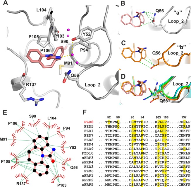
Figure 5.

Interactions between carbamazepine and FZD8CRD. (A) Details of the carbamazepine binding site (gray cartoon, from molecule “a” of the ASU, PDB code 6TFB). The carbamazepine interacting amino acid side chains are shown as gray sticks. Water molecules within the pocket are shown as magenta balls, carbamazepine as salmon sticks, and hydrogen bonds as yellow dashed lines. (B–D) Conformations of Q56 from two carbamazepine binding molecules of the ASU, “a” (gray, B) and “b” (brown, C), and superimposed with two representative molecules from apo structures, (green and cyan; D). Interactions are defined as distances between protein and carbamazepine of less than 3.9 Å. The hydrophobic interactions are shown as green dashed lines. (E) LIGPLOT25 of FZD8CRD–carbamazepine interactions. The green lines indicate hydrophobic interactions. Carbamazepine carbon atoms are shown as black, nitrogen as blue, and oxygen as red spheres. Only molecule “a” from the ASU is shown. (F) Sequence alignment of human FZDs and sFRPs, with carbamazepine interacting FZD8 residues and all matching residues highlighted. The residue numbers indicated are for mouse FZD8.
