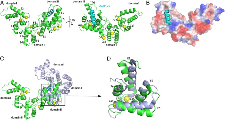
Fig. 2.
Crystal structure of drSCGN in complex with a SNAP-25 fragment and Ca2+. (A) Ribbon diagrams of drSCGN in complex with a SNAP-25 fragment, shown in two orientations rotated 180° with respect of each other. Green: drSCGN; cyan: SNAP-25 peptide; yellow balls: calcium ions. (B) Electrostatic potential surface of the complex of drSCGN with the SNAP-25 peptide (ribbon diagram). Blue: positive potential; red: negative potential. The complex is shown in the same orientation as that of the right molecule in A. (C) SCGN has different domain arrangement in the Apo (light blue) and Holo forms (green). The two structures were aligned by superimposing their domain III. (D) Overlay of domain III of SCGN in the Apo (light blue) and Holo forms (green).

