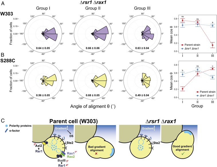
Fig. 7.
Improved gradient sensing in cueless (Δrsr1 Δrax1) cells. (A and B) Δrsr1 Δrax1 cells in the W303 (YGV5838) (A) and S288C (YDV6164) (B) backgrounds were exposed to an α-factor gradient generated in a microfluidic device, and the angles of the MPs relative to the gradient, θ, were measured. (Left) Data are shown in polar histograms as in Fig. 2C. Number of cells: 311 (YGV5838) and 390 (YDV6164). (Right) The means of cosθ ± SEM, indicated within the polar histograms, are plotted for the parental and Δrsr1 Δrax1 strains in both backgrounds. Data for parent strains are from Fig. 2C and SI Appendix, Fig. S2C. Statistical differences between parent and Δrsr1 Δrax1 were calculated by Kolmogorov–Smirnov test (P values: a, 3.5 × 10⁻⁷; b, 1.9 × 10⁻⁹; c, 0.049; ns, nonstatistical differences). (C) Model for the competition between internal cues and gradient decoding. In RSR1 RAX1 cells, the presence of internal cues can compete for a shared pool of polarity proteins, thereby affecting the cell’s ability to track the gradient. In the scheme, only competition for cytosolic components (blue circles) is represented, but the same argument can be applied for membrane components (Discussion). In Δrsr1 Δrax1 cells, the absence of internal cues removes the competition, improving the detection of gradient direction. In the parental W303 strain, the order of strength of the different landmarks is depicted by red numbers: Rax1-Rax2Distal > Axl2Proximal ≥ Bud8Distal > Rsr1(itself)Distal. (In S228C-background cells, Axl2 is stronger than Rax1-Rax2; see also SI Appendix, Table S2.)

