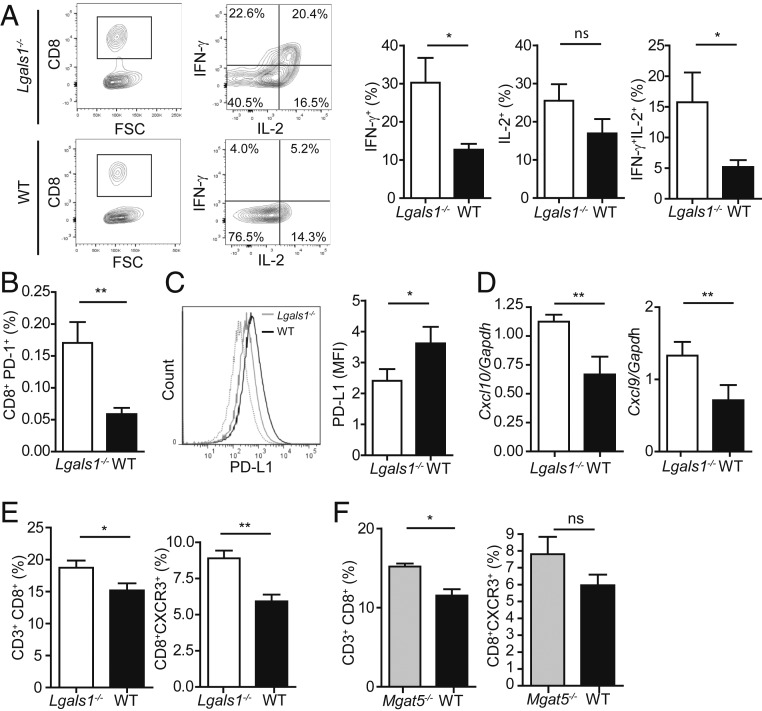
Fig. 3.
Gal1 deficiency enhances CD8+ T-cell function in salivary glands. (A) Cytokine production by salivary gland-infiltrating CD8+ T cells from aged Lgals1−/− and WT mice (9 mo). (Left) Representative dot plots and (Right) percentage of IFN-γ+, IL-2+, or IFN-γ+IL-2+ CD8+ T cell populations (mean ± SEM, n = 8). (B) Frequency of CD8+PD-1+ cells on salivary glands of aged (9 mo) Lgals1−/− and WT mice (mean ± SEM, n = 8; C). PD-L1 expression on salivary gland cells from aged Lgals1−/− and WT mice (9 mo; mean ± SEM; n = 8). (Right) Mean fluorescence intensity (MFI) and (Left) representative histogram. (D) Expression of Cxcl10 and Cxcl9 mRNA by RT-qPCR in salivary glands from aged Lgals1−/− and WT mice (9 mo; n = 10). Results are expressed relative to Gapdh mRNA expression. (E and F) Quantification of the frequency of CD8+ and CD8+CXCR3+ T cells in the submandibular lymph nodes (SLNs) of aged Lgals1−/−, Mgat5−/−, and WT mice (9 mo). Comparison between Lgals1−/− and WT mice (mean ± SEM, n = 16; E) and between Mgat5−/− and WT mice (mean ± SEM, n = 8; F). *P < 0.05; **P < 0.01; ns, nonsignificant; Student’s t test.

