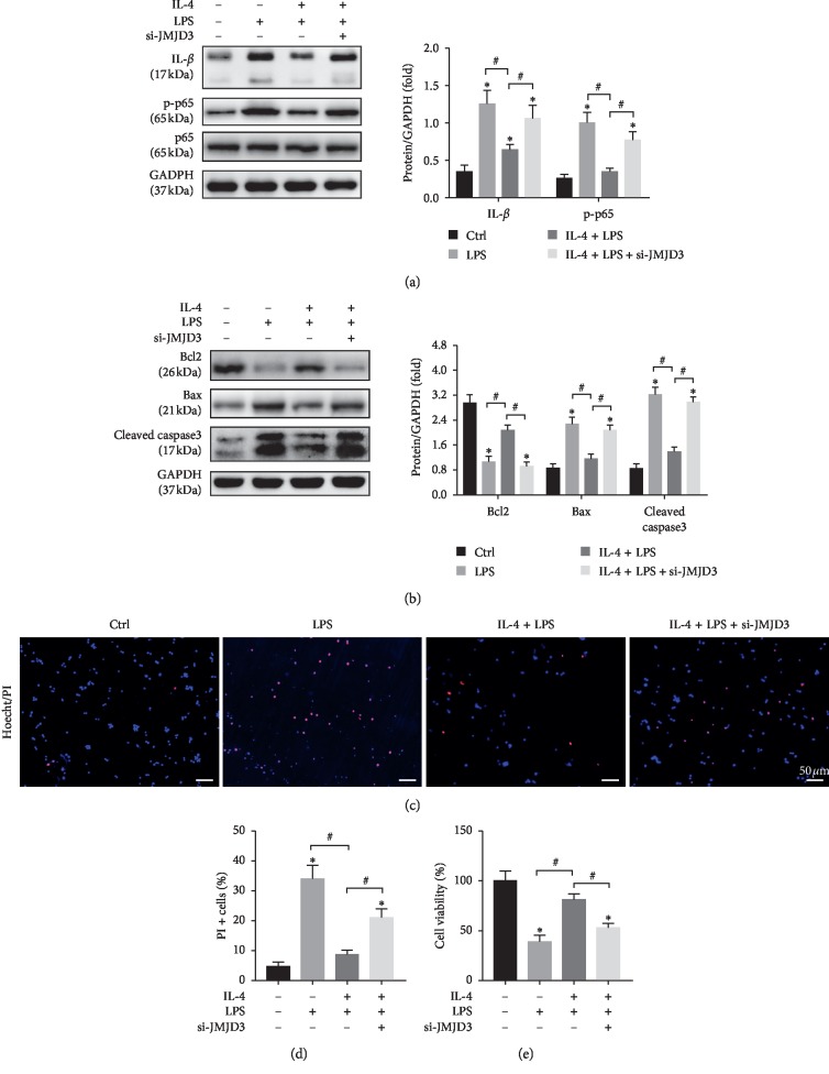
Figure 5.
JMJD3 knockdown weakened the anti-inflammatory and antiapoptotic effects of IL-4 in vitro. KCs and primary hepatocytes were isolated from untreated livers. KCs were treated with 100 ng/mL LPS for 24 h with or without treatment of 15 ng/mL IL-4 for 12 h The supernatant of the KCs was collected and used to stimulate primary hepatocytes. (a) The protein expression levels of IL-β, p-p65, p65, and GAPDH were detected by Western blot. (b) The protein expression levels of Bcl2, Bax, cleaved caspase3, and GAPDH were detected by Western blot (n = 3/group). GAPDH served as an internal control and was used for normalization (n = 3/group). (c) Representative images of Hoechst33342 staining for late apoptosis and necrotic cells: hoechst333424 (blue) and PI (red). (d) Quantification of PI-positive stained cells (n = 3/group). (e) Cell viability was detected by CCK8 (n = 3/group). The data are presented as the mean ± SD, ∗p < 0.05vs. the control group, #p < 0.05vs. the IL-4 + LPS group.

