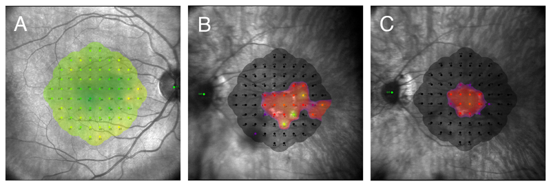
Figure 2.
Standard 10-2 grid consisting of 68 retinal points arranged in a Cartesian pattern covering the central 20°. The threshold sensitivity value at each retinal location is colour coded and shown as an overlay on the near-infrared image. A) Macular sensitivity heat map of a healthy 27 year-old male. Green indicates normal sensitivity (maximum 36dB). B and C) Macular sensitivity heat map of a 38 year-old male with RPGR X-linked RP taken two years apart. In 2017 (B) the patient still showed few retinal points of nearly normal sensitivity, while two years later (C) both the central visual field constriction, and sensitivity threshold had worsened.

