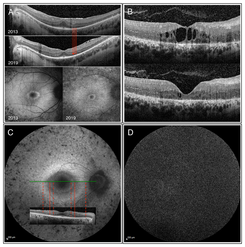
Figure 4.
OCT and autofluorescence images of various patients with RP. A) Images of the right eye of a 28 year-old male with autosomal recessive RP (compound heterozygous mutations in DFNB31 and USH2A found). Significant decrease in the width of both the ELM and EZ paralleled by diminishing foveal hypoautofluorescence can be observed between 2013 and 2019. Visual acuity in 2019 was 6/7.5, which translates to 20/25 (20 ft) or 0.8 (decimal) Snellen equivalent. B) OCT images of the right eye of a 26 year-old female patient with CRB1 associated RP showing clinically meaningful improvement of the cystoid macular oedema upon systemic acetazolamide therapy with an increase in visual acuity from 3/60 (20/400, 0.05) to 6/38 (20/125, 0.16). C) Autofluorescence and OCT images of the right eye of a 61 year-old male with PRPF8 associated autosomal dominant RP. The inner hyperautofluorescent circle shows good correlation with the loss in ELM, while the outer larger hyperautofluorescent ring correlates with retinal area of absent ELM and highly thinned ONL. D) Short-wavelength autofluorescence image of a 48 y/o male patient with biallelic RPE65 (c.11+5G>A, c.1543C>T) showing the characteristic absent autofluorescence.

