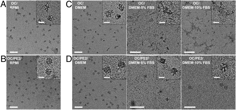
Fig. 6.
The effect of peptoid coating of OCs in cell media and presence of serum nuclease. TEM images show bare OCs (A and C) and PE2-coated OCs (OC/PE2) (B and D) in RPMI (A and B) and DMEM (C and D) containing 0%, 5%, and 10% FBS and incubated at 37 °C for 24 h. The final concentrations of MgCl2 were 1.25 mM. TEM imaging was performed on samples extracted from the agarose gels. (Scale bars, 200 nm.) A–D, Insets show magnified images of the OC structures. (Scale bars, 50 nm.)

