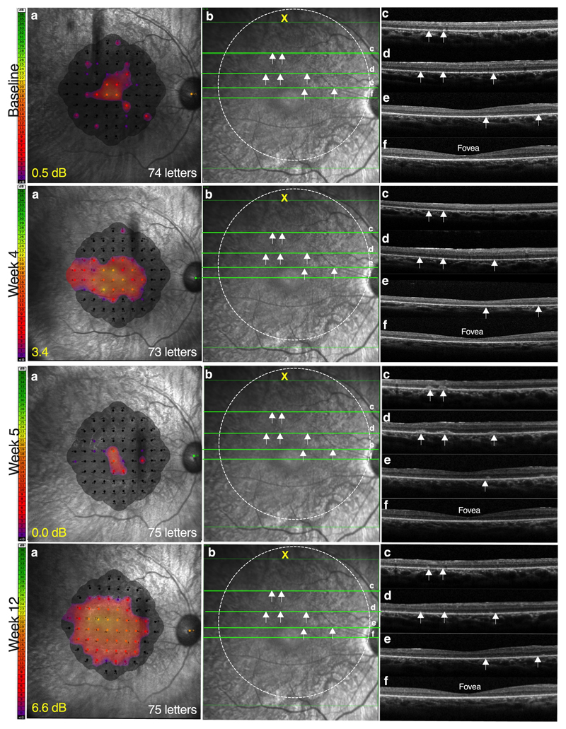
Fig. 2. Transient regression of retinal function in the treated eye associated with subretinal inflammation at 5 weeks post high-dose gene therapy.
At week 5 post treatment with AAV8.coRPGR (6x10¹⁰ gp), patient C4.1 noticed some regression of vision in the treated eye following improvements over the preceding week. Microperimetry showed regression of retinal sensitivity and visual field (a), which was associated with subretinal lesions (white arrows) on optical coherence tomography (OCT) (b). Subretinal lesions (white arrows) could be seen to be scattered over the treated area of the macula on the OCT cross-sections (c-e), but spared the fovea (f); visual acuity was unaffected following treatment with a course of oral corticosteroids, the inflammation resolved with corresponding gains in retinal sensitivity recorded at 3 months follow-up.

