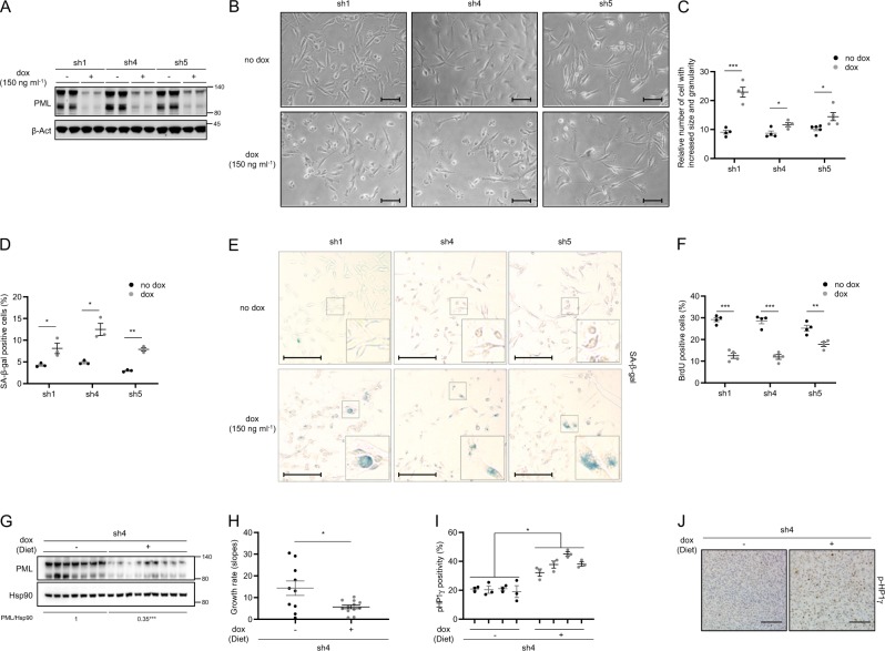
Fig. 1.
PML silencing induces senescence. Effect of doxycycline-inducible (150 ng ml−1; 3 + 3 days) PML silencing (sh1, sh4, and sh5) on PML protein expression (a, representative of at least three experiments), on the morphology (b, representative images, scale bar, 50 μm), on cell size and granularity (c, FACS analysis, sh1 and sh4, n = 4, sh5, n = 5), on the number of senescent cells (d; n = 3, representative images of SA-β-Galactosidase assay, scale bar 50 μm (e)) and on the number of BrdU positive cells (f, n = 4) in MDA-MB-231 cells. Impact of doxycycline-inducible PML silencing (sh4) of established MDA-MB-231 xenografts on PML protein expression (g), on tumor growth rate represented as the growth rate of each tumor (h, sh4 no dox, n = 10; sh4 dox, n = 12; growth rate was inferred from the linear regression calculated for the progressive change in tumor volume of each individual tumor during the period depicted in Supplementary Fig. 1p) and on number of senescent cells measured by p-HP1γ staining (i, sh4 no dox, n = 4; sh4 dox, n = 4); representative images of p-HP1γ positive cells, scale bar 100 μm (j) of the tumors. Error bars represent s.e.m. p, p-value (*p < 0.05, **p < 0.01, ***p < 0.001). One-tailed Student's t-test was used for cell line data analysis (c, d, f) and one-tailed Mann–Whitney U-test for xenografts (h, i). sh1, sh4, and sh5: shRNA against PML, dox: doxycycline, SA-β-gal: senescence-associated beta-galactosidase, BrdU: bromodeoxyuridine, p-HP1γ: phospho-heterochromatin protein-1 gamma, molecular weight markers (kDa) are shown to the right

